
An echocardiogram is a noninvasive imaging test that uses ultrasound waves to create detailed pictures of the heart. It allows providers to assess heart structure, function, and blood flow without radiation or contrast agents. This test is safe and widely used for diagnosing various heart conditions. The echocardiogram can provide real-time images, helping clinicians evaluate heart valves, chambers, and muscle thickness. Because it is painless and accessible, it often serves as a first-line diagnostic tool in cardiology.
How Does an Echocardiogram Work?
The procedure involves placing a transducer on the chest to send and receive sound waves. These waves bounce off heart tissues, producing echoes that a computer converts into images. The technician guides the transducer to capture views from multiple angles, providing a comprehensive evaluation. Doppler technology within the echocardiogram measures the speed and direction of blood flow through the heart’s chambers and valves. This information helps detect abnormalities such as valve leaks or blood flow obstructions.
What Conditions Can Echocardiograms Diagnose?
Echocardiograms diagnose a range of heart diseases and structural abnormalities. They detect valve disorders such as stenosis or regurgitation, cardiomyopathies, congenital heart defects, and pericardial effusion. The test can also identify decreased heart pumping function, heart muscle thickening, or fluid buildup around the heart.
In patients with symptoms like chest pain, shortness of breath, or palpitations, an echocardiogram helps pinpoint the underlying cardiac cause. It also guides treatment decisions for heart failure and arrhythmias. Providers recommend echocardiograms when patients present with signs of heart disease or have risk factors like hypertension or diabetes. It is useful for evaluating murmurs heard during physical exams or monitoring known cardiac conditions.
The test is commonly ordered before and after cardiac surgeries or interventions to assess heart function. Echocardiograms may also support routine screenings in high-risk individuals or follow-up care after heart attacks. Timing depends on clinical need and symptoms.
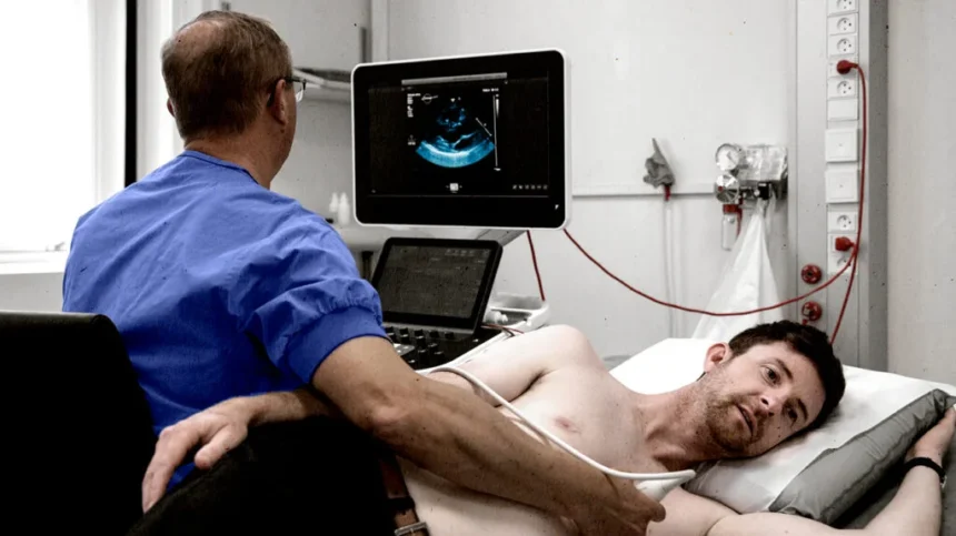
How Is the Procedure Performed?
During the test, patients lie on an exam table while the technician applies gel to the chest area. The transducer moves gently across the skin to capture images, typically taking 30 to 60 minutes. The procedure is painless and requires no special preparation, allowing patients to resume normal activities immediately afterward. In some cases, transesophageal echocardiography may be performed, involving a small probe inserted into the esophagus for clearer images of certain heart structures. The choice depends on the clinical question.
Echocardiogram results provide detailed information about heart size, wall motion, valve function, and blood flow patterns. They help identify weak or damaged areas of the heart muscle and quantify ejection fraction, a measure of pumping efficiency. Abnormal findings may indicate the need for further testing or guide medication adjustments. Results also help stratify patient risk and guide lifestyle or procedural interventions. The clarity and precision of echocardiogram data contribute to personalized cardiac care.
Discuss an Echocardiogram
If you experience symptoms such as chest discomfort, fatigue, or irregular heartbeat, consider discussing an echocardiogram with your healthcare provider. Those with known heart conditions or multiple cardiovascular risk factors may also benefit from regular imaging to monitor heart function. Early evaluation through echocardiography can facilitate timely diagnosis and tailored treatment. Ask your cardiologist how an echocardiogram fits into your overall heart health plan and what to expect from the procedure.