
An echocardiogram is a noninvasive test that uses sound waves to create detailed images of your heart, helping doctors assess its structure and function. Whether it’s part of a routine check-up or ordered to investigate specific symptoms, understanding what to expect can help you prepare for the procedure. Here’s what you need to know about preparing for your echocardiogram:
Understanding Echocardiograms
An echocardiogram, often called an echo test or heart ultrasound, uses sound waves to create moving pictures of your heart. The test measures the size and shape of your heart, assesses how your heart chambers and valves function, and evaluates the flow of blood through your heart. Doctors order echocardiograms for several reasons. They use the test to detect heart disease, measure how well your heart pumps blood, and identify problems with heart valves. It can also check for blood clots inside your heart or find fluid around your heart.
Preparing for Your Echocardiogram
Most echocardiograms require little to no preparation. Follow these guidelines to prepare for your test:
- Wear comfortable, loose-fitting clothing to your appointment.
- Be prepared to remove your shirt so the technician can place sensors on your chest. A medical gown will be provided for you to wear during the test.
- Inform your doctor about any medical devices, such as a pacemaker, that you have.
- Mention any allergies or if you are pregnant, as this information helps your doctor plan the safest approach.
- If you are undergoing a transesophageal echocardiogram, you may need to refrain from eating or drinking for several hours beforehand. Your doctor will provide specific instructions in this case.
Following these steps helps you with a smooth and efficient experience for your echocardiogram.
Knowing What To Expect
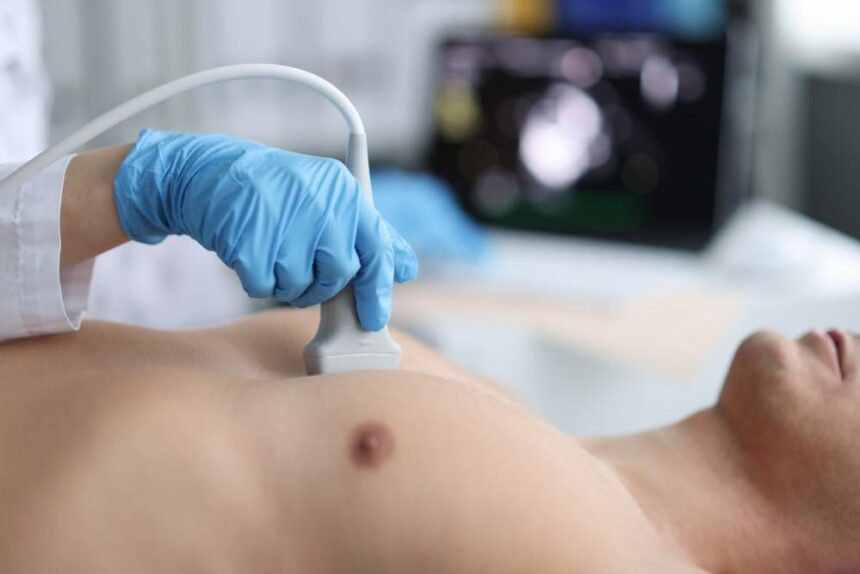
A trained technician, called a cardiac sonographer, performs the test. You’ll lie on an exam table, usually on your left side, and the technician will attach small sensors called electrodes to your chest. These sensors track your heart rhythm during the test. Next, the technician will apply a clear gel to your chest. This gel helps sound waves travel better.
The technician will then move a small device called a transducer across your chest. This device sends sound waves through your chest wall to your heart. The sound waves bounce back and create pictures on a computer screen. You may hear whooshing sounds during the test; these are the sounds of blood flowing through your heart. The test doesn’t hurt, but may might feel some pressure when the technician presses the transducer against your chest. You may also need to hold your breath briefly or change positions so the technician can get clear pictures from different angles.
Reviewing and Interpreting Results
Your results won’t be ready right away. A cardiologist will review the images and prepare a report, which usually takes a few days. The report includes measurements of your heart chambers and an assessment of your heart’s pumping ability.
The report also describes your heart valves and checks whether they’re opening and closing properly. It detects any abnormal blood flow patterns and measures the pressure in various parts of your heart.
Your doctor will explain what the results mean for your health. Some findings may indicate that your heart is functioning normally, while others may reveal problems that require treatment or further testing. Your doctor will discuss any follow-up steps based on your results.
Schedule your echocardiogram today
An echocardiogram is a helpful tool for checking your heart health. The test is quick, safe, and provides detailed information about how your heart works. Most people require little preparation and can return to their normal activities immediately after the test. If your doctor has recommended an echocardiogram, contact a cardiologist near you to schedule your appointment today.